
Case Studies By Dr. Glossy
*Disclaimer - Content displayed below, under ‘case studies’ is for medical professionals. Published for educational and informational purposes only and is not intended as a diagnosis, treatment or as a substitute for professional medical advice, diagnosis and treatment. Certain pictures might not be suitable for others or non-medical professionals.
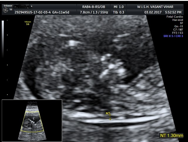
NT SCAN criteria
NT SCAN criteria according to the ISUOG -International society of Ultrasound and Obstetrics and Gynecology and FMF- fetal medicine foundation, UK criteria
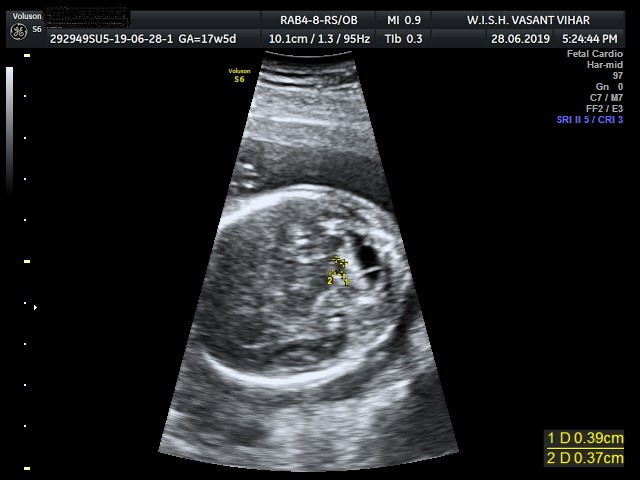
Fourth ventricle Index (4VI)
Fourth ventricle Index (4VI): A sonographic marker for severe fetal vermian dysgenesis/agenesis
Intracranial translucency
Intracranial translucency as a sonographic marker for detecting open spina bifida (OSB) at 11–13 +6 weeks scan
Iniencephaly Apertus
Ultrasound Star Gazing fetus- ‘Iniencephaly Apertus’
NT SCAN criteria
NT SCAN criteria according to the ISUOG -International society of Ultrasound and Obstetrics and Gynecology and FMF- fetal medicine foundation, UK criteria
- Gestational period must be 11–13 weeks and six days.
- The fetal crown-rump length (CRL) should be between 45 and 84 mm
- Magnification- to include only fetal head and upper thorax.
- True mid sagittal plane- defined by the presence of the echogenic tip of the nose and rectangular shape of the palate anteriorly, the translucent diencephalon in the center and the nuchal membrane posteriorly
- Neutral fetal position- In order to ensure that the fetus is not flexed, amniotic fluid should be visible between the fetal chin and chest
- Calipers “ON to ON”— The ultrasound machine should allow measurement precision of 0.1 mm. Calipers should be placed correctly (on-on) to measure NT as the maximum distance between the nuchal membrane and the edge of the soft tissue overlying the cervical spine.
- Maximum lucency to be taken- If more than one measurement meeting all the criteria is obtained, the maximum one should be recorded and used for risk assessment
- The nuchal membrane must be seen
For further information please email us at [email protected]
Fourth ventricle Index (4VI)
Fourth ventricle Index (4VI): A sonographic marker for severe fetal vermian dysgenesis/agenesis.
- Although fourth ventricle is not part of the routine assessment as recommended by current ISUOG guidelines, “open 4V” is a sonographic marker seen in the second trimester and is a indicator of MB-HB (midbrain-hindbrain) malformations.
- Measure the fourth ventricle in the APD(anteroposterior) and LLD (laterolateral) dimensions in posterior fossa view. 4VI=LLD/APD.
- A 4VI below -2SD from the mean or 4VI < 1 indicates the need for a detailed study of the MB-HB structures.
Ref: Fourth ventricle Index : A sonographic marker for severe fetal vermian dysgenesis/agenesis: Ultrasound Obstet Gynecol 2019; 53:390-395- Published online 31 January 2019 in Wiley Online Library (wileyonlinelibrary.com). DOI: 10.1002/uog.19034
For further information please email us at [email protected]
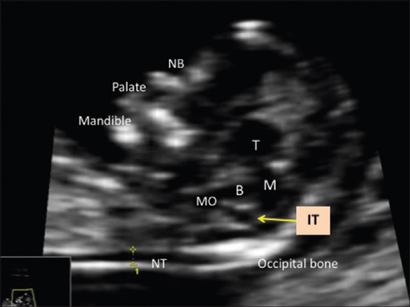
Intracranial translucency
Intracranial translucency as a sonographic marker for detecting open spina bifida (OSB) at 11–13 +6 weeks scan:
- Mid-sagittal view of face that is routinely used to measure nuchal translucency (NT) can also be used to detect OSB. It is feasible to integrate IT into the routine 11–13 +6 weeks scan.
- Diagnosis of open spina bifida (OSB) in the antenatal period is routinely done in the mid-trimester using lemon and banana signs on ultrasound. Fourth ventricle is seen as intracranial translucency (IT) between 11 and 13 +6 weeks of gestation. Obliterated IT has been identified as a marker for ONTD
- IT is identified in the mid-sagittal image of fetal face as a clear space bounded anteriorly by posterior border of brainstem and posteriorly by choroid plexus of fourth ventricle.
Ref: IJRI journal- year 2017, volume 27, issue4, page 427 431: Intracranial transluceny as a sonographic marker for detecting open spina bifida at 11-13+6 weeks scan: Our experience: Madhavi L Teegala, Dhamangaonkar G Vinayak
For further information please email us at [email protected]
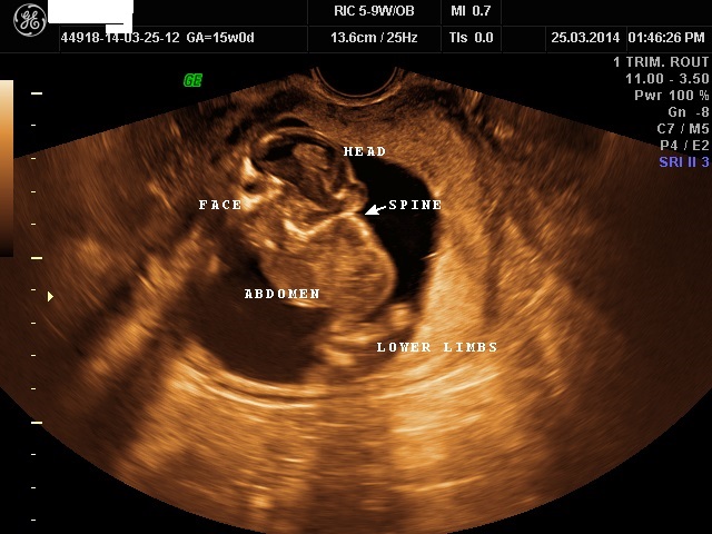
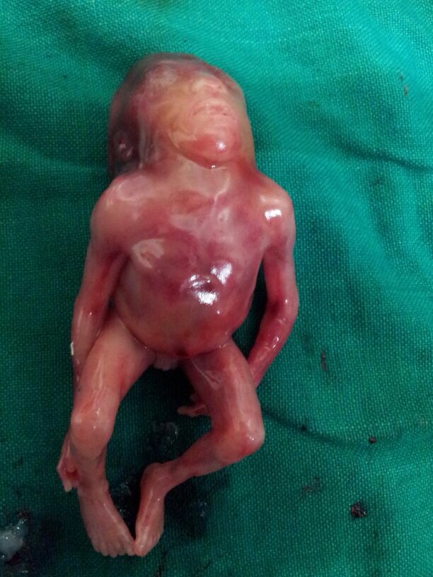
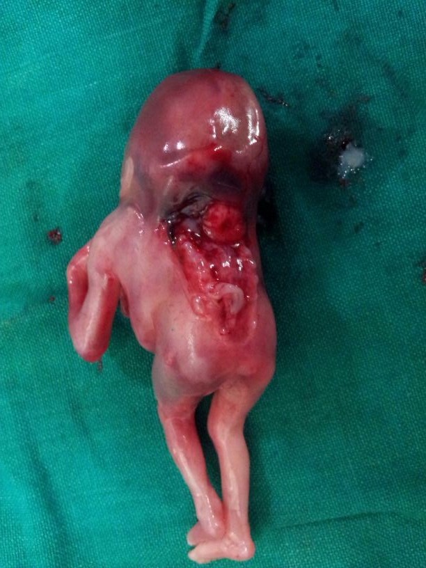
Iniencephaly Apertus
Ultrasound Star Gazing fetus- ‘Iniencephaly Apertus’
- Iniencephaly is a rare neural tube defect characterized by extreme retroflexion of the head with absence of the neck due to spinal deformities.
- On antenatal ultrasound scan, features that clinch the diagnosis of iniencephaly
i. deficit of occipital bone with an enlarged foramen magnum
ii. fusion of malformed cervical and thoracic vertebrae
iii. upward turned face with chin being continuous with the chest because of absence of neck. - Our patient- A 33-year-old multigravida woman visited our department for routine obstetric check up at 15 weeks of gestation. She had neither had prior antenatal check up nor folic acid supplementation. She had been a hypothyroid for 4 years and was on thyroxine supplementation (50 mcg once daily). She did not have any consanguine relationship with her husband. There was no history of teratogen exposure.
- An antenatal diagnosis of inencephaly apertus was made. In view of obvious fetal anomalies incompatible with extra-uterine life, the parents were counselled for MTP. Postnatal correlation suggested the same.
For further information please email us at [email protected]
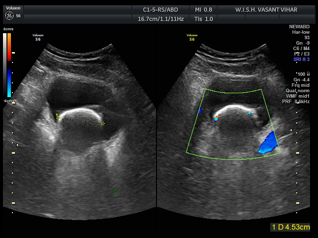
Vesical calculus
- Either primary (origin in urinary bladder) or secondary (from renal origin through ureter)
- Usually associated with urinary stasis and Common in males
- Surgery is the treatment of choice for large vesical calculus
- In our case below, male child 10 years old shows a large single mm vesical calculus. There was history of recurrent urinary tract infections along with nutritional deficiencies (Vitamin A and Vitamin B6, low protein and high carbohydrate diet). Surgical removal was done and child had normal post-operative course.
For further information please email us at [email protected]
“My vision is to spread awareness; to support and serve everyone in recognising that health is their most valuable asset.”
Dr. Glossy Sabharwal
MBBS, MD Radiology
Fellowship in Gynaecology
Fellowship in Breast Imaging
Harvard University & Yale School of Medicine Certified
Director & Head of Radiology, Jeewan Mala Hospital, JMH Prime hospital and WISH Clinic